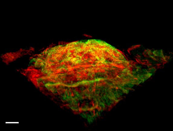
The intestinal landscape
Affiliation:
ESRIC
Description:
The image shows a section of mouse intestine, with each cell stained with a green dye for DNA and a red dye for the filamentous structure actin. The scale bar is 5 μm. The image is made up of a stack of images which have been taken moving through the thick tissue section.
Equipment used:
Leica Microsystems SP5 SMD gated-STED microscope (laser scanning confocal microscope)